A Case of Intramuscular Lipoma Arising in the Inferior Surface of the Tongue
2Department of Collaborative oral health (Oral Radiology), Matsumoto Dental University Hospital, Nagano, Japan
3Department of Oral pathology, School of Dentistry Matsumoto Dental University, Nagano, Japan
4Department of Oral Health Promotion, Graduate School of Oral Medicine, Matsumoto Dental University, Nagano, Japan
5Department of Oral and Maxillofacial Surgery, School of Dentistry Matsumoto Dental University, Nagano, Japan
Intramuscular lipoma is a benign soft tissue tumor that accounts for 1.8 to 2% of all lipomas, which often originates from the large muscles in the extremities, rarely in the oral region. This time, we experienced a case of intramuscular lipoma on the undersurface of the left side of the tongue and reported the overview with literal consideration. The patient was a 62-year-old male, and complained of indolent swelling on the undersurface of the left side of the tongue and sought medical attention. Relatively well-demarcated low-density area approximately 10 mm in greatest dimension was observed in the left side of the tongue tip on the CT images, while relatively well-demarcated area with hyperintensities in the inside and partially muscular tissues and partially linear portion showing the same signal with muscular tissues on T1- and T2-weighted MRI images. A diagnosis of lipoma was made based on clinical findings and imaging findings of the CT and MRI, and removal was performed using general anesthesia. Histopathologically, the fat tissues had no surrounding membranes and were lobulated and grew into the muscular layer, tumor cells were large, adipocyte-like cells with clear cytoplasm grew into the muscular layer, and a diagnosis of intramuscular lipoma was made. We searched for and collected case reports on intramuscular lipoma in the stomatognathic area in Japan and identified 14 cases including this case. Particularly, 3 cases of intramuscular lipoma on the undersurface of the tongue including this case were identified and the location was considered to be rare. Recurrence rate of intramuscular lipoma was high level of 3% to 62.5%, and it is important to examine much imaging information in detail from imaging diagnosis with CT and MRI examinations. This patient was well after surgery without signs of recurrence.
Conflict of interest and Ethical prospect
In this article, we do not have any conflict of interest (COI). As for ethical prospect, we have complied with the Declaration of Helsinki and have obtained informed consent from the patient for this case report. This article was disclosed partially at the 7th Clinico-pathological conference (CPC) at Matsumoto Dental University Hospital on February 19, 2015.
Introduction
Lipoma is the most commonly-observed nonepithelial benign tumor among soft tissue tumors which consist of mature adipose cells and develop subcutaneously. Lipoma is categorized to superficial lipoma which is observed in the subcutaneous tissues and to deep lipoma which is observed in the subfascial, intramuscular and intermuscular tissues. Mostly it is a soft single tumor which consists of mature adipose tissue, while it becomes multiple in a rare frequency. It likely develops in the subcutaneous tissue of back, shoulder, face, neck and thighs. Intramuscular lipoma is a benign soft tissue tumor which accounts for 1.8 to 2% of whole types of lipoma[1]. It likely occurs in the muscles of extremities and rarely occurs in the oral area. At this time, we experienced a case of single intramuscular lipoma which occurred in the left sublingual region; thus report the summary with the imaging and by adding some bibliographic consideration.
A 62-year-old male patient found painless swelling in the sublingual region from the beginning of September 2014, which was disturbing; thus visited a nearby dentist then our hospital for a work-up. His general physical findings on the first visit were body height: 165 cm, body weight: 70.4 kg, body temperature: 35.1°C, and blood pressure: 145/85 mmHg. The local appearance was a pale-yellow, soft-elastic painless bump of 20 mm in longitudinal axis x 15 mm in horizontal axis in the left inferior surface of the tongue. No redness and haphalgesia was observed in the mucosa of that area, and nothing was palpable in the neck and the submandibular lymph nodes. His medical history was diabetes mellitus (HbA1c: 6.9, blood glucose level: 143) and hypertension since the age of 28-year-old, for which he has been treated with pioglitazone hydrochloride, sitagliptin phosphate hydrate and voglibose and has been on regular visits for the treatment. There was no remarkable family history. Due to swelling observed in the left sublingual region at the consultation, CT and MR tests were performed for a work-up. In the CT imaging, a low density area with the greatest dimension of approximately 10 mm with relatively clear borders was found in the left proglossis part, and the internal CT value indicated -108 (Fig 1A, B). In the MR imaging, a round-like shaped tumor mass formation figure of around 1 cm dimension was observed in the slightly left area on the inferior/center of proglossis part, and a high signal area was found inside with relatively clear borders and a linear site which showed partially the equal signal to the muscular tissue was found in the T1-highlighted imaging (Fig 2 A, B) and T2-highlighted imaging (Fig 2 C, D). A low signal area was found in the fat-restraining T2-highlighted imaging (Fig 2 E, F).

(B) The internal CT value indicated -100, suggesting lipid (arrow).

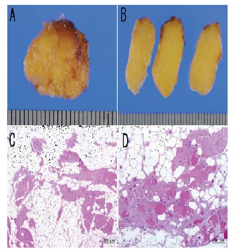
Suspected lipoma in the sublingual region was diagnosed according to the clinical diagnosis, CT and MR imaging diagnosis. Tumor mass enucleation was performed under local anesthesia in late October 2014. After peeling off the covering mucosa then confirming the adipose tissue, the tumor mass was enucleated as one mass by bluntly peeling off from the surrounding tissue. The macroscopical findings of the enucleated part from the left sublingual region were pale yellow, smooth on the surface and soft-elastic (Fig 3 A, B). In the histopathological diagnosis, the tumor mass consisted of adipose tissue without clear surrounding capsular structure. This adipose tissue was separated lobularly with the fibrous tissue with observed proliferation into the muscular wall (Fig 3 C). The adipose tissue which composed the tumor was big and mature adipocyte, with big and clear cytoplasm, and with the nuclear compressed to the cellular rim (Fig 3 D). While these tumor cells were mildly different in size, it was not observed to be clearly atypical. In addition, no lipoblast-like cells were observed within the tumor tissue. The histopathological diagnosis was intramuscular lipoma. The healing process of the surgical wound was good, and the patient has been under regular outpatient observation by explaining that sufficient observation is required.

Discussion
Lipoma is a mesodermal, nonepithelial benign tumor which consists of mature adipose tissue, and has been reported to be one of the most frequent benign tumors[1, 2]. Lipoma grows relatively slowly to form painless swelling, which is palpable as a movable soft orbicular or lobular tumor mass with relatively clear borders.
Histopathological classifications include, according to the substrate and appearance of tumor cells, simple lipoma, fibrous lipoma, myxomatous lipoma, vascular lipoma, spindle cellular lipoma, polygonal lipoma, parosteal lipoma and muscular lipoma[1-4]. With regard to histopathological incidence of lipoma in the mouth, Oku et al[5] reported that simple lipoma is the most frequent with 62.2% (123/201 patients) followed by fibrous lipoma with 35.8% (72/201 patients), and these two types accounted for most of the cases. Muscular lipoma develops within the muscular tissue and grows due to infiltrating mature adipose cells, which are tumor cells, deep into the muscle fibers. Histopathologically, it is characteristic that existing muscle fiber bundle remains within the tumor. Pélissier et al.[6]reported about the incidence of intramuscular lipoma, that 4 out of 7 cases of intramuscular lipoma which developed in the mouth were on the tongue. In Japan, Kobayashi et al.[7] reported 6 cases of intramuscular lipoma which developed on the tongue. In addition, Enjoji et al.[3] reported that 27 cases of intramuscular lipoma (1.7%) were included in the 1615 cases of lipoma; thus, the incidence is considered “rare”.
At this time we newly found 14 cases including self-experiments[7-17]in the search and found by Pub Med, CiNii Article for case reports of intramuscular lipoma which developed on the tongue in the stomatognathic area (excluding reports at academic conferences) in Japan (Table 1). In addition, while some of the searched cases were described as infiltrating or intramuscular lipoma, they were categorized into intramuscular lipoma according to the Shuman[18]classification. The age was within 37 to 75 year-old and the mean age was 62 year-old. Since there are reports that lipoma in the oral area frequently occurs in the age range of 30 to 60 year-old and it is observed at the various age of 1 to 72 year-old[19], the difference in age is not consistent among reports. However, lipoma mostly occurs in middle age to elderly people, which was not significantly different from the age for frequent occurrence of simple lipoma. With regard to the gender difference for lipoma, while Kobayashi et al.[7]in Japan reported their determination with regard to glossal lipoma of the trend that it occurred more frequently in male (39 male subjects vs. 16 female subjects), no significant difference was observed for intramuscular lipoma which developed on the tongue (7 male subjects vs. 7 female subjects) in our determination. With regard to clinical symptoms, most of them are painless tumor mass and swelling on the tongue, followed by less frequent articulatory disorder, lodging sensation of pharynx, and ear pain, which are observed to be normal clinical symptoms of lipoma and no specific clinical symptoms for intramuscular lipoma may exist. With regard to the onset site, onset in the glossal edge part was the most common with 10 cases. Those which developed in the inferior surface of the tongue were in 3 cases including self experiments; thus it is considered rare onset site. With regard to the size, while they were 50 to 7 mm in size and no specific size was observed, given that intramuscular lipoma is reported to be found mostly when they grow to a certain size compared to simple lipoma and intramuscular lipoma was reported to unlikely show manifestations due to its deeper location and slower growth than simple lipoma by Piattelli et al.[20], no specific subjective symptoms were observed until the patient her/himself got aware in the self experiments as well.

With regard to the mechanism of development of lipoma, while there are hypotheses such as ectopic development theory[21], and hyperplasia theory of normal tissue[22], the causes such as inherited predisposition, congenital intrinsic factor, endocrine deconditioning, impaired metabolism of lipid, trauma, and chronic stimulation[23] have been reported; however, the causality has not been currently clarified. In the self experiments, the medical history included diabetes mellitus and hypertension, the causes could not be confirmed.
Lipoma is categorized to superficial lipoma which is observed in the subcutaneous tissues and to deep lipoma which is observed in the subfascial, intramuscular and intermuscular tissues[24]. While superficial lipoma is relatively easy to be diagnosed due to its likely clearer swelling compared to deep lipoma, deep lipoma may be difficult to be diagnosed depending on its onset site; thus, CT and MR tests are very useful imaging tests for the diagnosis of lipoma. It is reported specific for intramuscular lipoma to have a CT imaging finding of a high density linear figure which is assumed to be residue muscular tissue in the tumor showing a uniform lipid concentration[25,26]. In addition, it has been reported that some irregular parts in the low-density tumor mass with clear borders, which indicated equal density as the muscular layer, were observed in contrast CT imaging[7]. While we did not perform a contrast CT test for this patient, a high density linear figure which is assumed to be residue muscular tissue in the tumor was shown in the simple CT imaging finding. With regard to the MR imaging findings, it has been reported that equal signal parts to the muscular tissue were mixed with unclear borders in the same high signal area as adipose tissue in the both T1 and T2-highlighted imaging, and that linear figures with equal signal to muscles were partially observed in the T2-highlighted imaging coronal section[27]. In this case, MR findings showed the tumor mass with clear borders in both T1-highlighted imaging and T2-highlighted imaging, with internal area mostly shown as a high signal, and parts of equal signal to the muscular tissue mixed with unclear borders and linear sites with equal signal to the muscular tissue were observed inside. While it is considered that our case at this time showed imaging findings which are characteristic for intramuscular lipoma, it is essential for a final diagnosis to determine by a histopathological search.
References:
- Fletcher CD, Martin-Bates E. Intramuscular and intermuscular lipomaneglected diagnoses. Histopathology 1988; 12: 275-87.
- MacGregor AJ , Dyson DP. Oral lipoma. A review of the literature and report of twelve newcases. Oral Surg Oral Med Oral Pathol 1966; 21: 770-77.
- Enjoji M, Iwasaki H, Komatsu K. Benign Soft-Tissue Tumors in Japan –A Statistic Analysis of 8,086 Tumors. Japanese journal of cancer clinics 1974; 20: 594-609.
- Epivatianos A, Markopoulos AK, Papanayotou P. Benign tumors of adipose tissue of the oral cavity: a clinicopathologic study of 13 cases. J Oral Maxillofac Surg 2000; 58: 1113-1117.
- Oku Y, Tanaka A, Watanaba Y, Suka N, Tanaka S, Ehara M, Araki H, Kusama K and Sakashita H. Tow case of intraoral lipoma. J Meikai Med 2007; 36: 227-232.
- Pélissier A, Sawaf MH, Shabana AH. Infiltrating (intramuscular) benign lipoma of the head and neck. J Oral Maxillofac Surg 1991; 49: 1231-1236.
- Kobayashi D, Kida A, Ogawa S, Kawamoto A, Yamauchi Y, Endo S , Sugitani M. A case of tongue lipoma. Pract otol 2002; 95: 365-370.
- Nakayama Y, Nishijima K, Nabeyama H, Kayano T, Ikegami N, Kuwana S, Nagai N.A case of bilateral infiltrating lipoma of the tongue. Jpn J Oral Maxillofac Surg 1986; 32: 1068-1073.
- Saka M, Shirasuna K, Kogo M, Watatani K, Sugiyama M and Matuya T. Intramuscular lipoma of the tongue. Jpn J Oral Maxillofac Surg 1988; 34: 1433-1436.
- Kanekawa A. A case of benign symmetric lipomatosis of the tongue. Jpn J Oral Maxillofac Surg 1989; 35: 1535-1537.
- Takeda Y. Intramuscular lipoma of the tongue: report of a rare case. Ann Dent 1989; 48:22-24.
- Hiramatsu M, Takahashi S, Kato I, Mukai H. Diseases of the tongue - mainly neoplasm. Lipoma. Pract Dermatol 1996; 18: 215-218.
- Nakano T, Aiba T, Kubo T, Yamada K, Wada K, Wada T, Uyama T. A case of intramuscular lipoma of the tongue. Otolaryngology-Head and Neck Surgery 2002; 74:953-955.
- Tanabe S, Hasegawa M, Kouno N. Intramuscular (infiltrating) lipoma of the tongue. Stomato pharyngol 2006; 18: 453-457.
- Naruse T, Yanamoto S, Kawano T, Yoshitomi I, Yamada S, Kawaseki G, Fujita S, Ikeda T and Umeda M. Intramuscular lipoma of the tongue: Report of case complicated with diffuse lipomatosis. J Oral Maxillofac Surg Med Pathol 2012; 24:237-40.
- Kamatani T, Kutsuna T, Yasuda A, Yoshiba S, Saito Y and Shintani S. A case of intramuscular lipoma arising in the inferior surface of the tongue. Jpn J Oral Maxillofac Surg 2014; 60: 291-294.
- Kinukawa M, Ueki T, Yoshida Y, Nagai S. A case of intramuscular lipoma arising from both borders of tongue. J J M C P 2014; 23: 90-94.
- Shuman R, Anderson WAD. Pathology. 7th ed. pp1888-1892. C. V. Mosby, St. Louis, 1977.
- Shizuku H, Kashima K, Sato G. A Case of Cervical Lipoma. Tokushima Red Cross Hospital Medical Journa 2002; l7: 66-68.
- Piattelli A, Fioroni M, Lezzi G, Rubini C. Osteolipoma of thetongue. Oral Oncol 2001; 37: 468-470.
- Blake H, Blake FS. Lipoma in the floor of the mouth ; report of a case. Oral Surg Oral Med Oral Pathol 1959; 12: 1436-1438.
- Stout AP. Liposarcoma-the Malignant Tumor of Lipoblasts. Ann Surg 1944; 119:86-107.
- Allard RHB, Blok P, van der Kwast WAM, van der Waal. Oral lipomas with osseous and chondrous metaplasia; report of two cases. J Oral Pathol 1982; 11:18-25.
- Kinoshita H, Ogasawara T, Hirai R. A case of a lipoma in the outer region of the angle of the mandible. Jpn J Oral Diag Oral Med 2013; 26: 222-225.
- Matsumoto K, Hukuda S, Ishizawa M, Chano T, Okabe H. MRI findings in intramuscular lipomas. Skeletal Radiol 1999; 28: 145-152.
- Ootsuji T, Shikata J, Kuzuoka T, Takahashi M, Kin E. An unusual variant of intramuscular lipoma in the tensor fascia lata. Orthop Surg Traumatol 1991; 34:1229-1231.
- Ohguri T, Aoki T, Hisaoka M, Watanabe H, Nakamura K, HashimotoH, Nakamura T, Nakata H. Differential diagnosis of benign peripheral lipoma from well-differentiated liposarcoma on MR imaging: is comparison of margins and internal characteristics useful? ARJ 2003; 180: 1689-1694.
Information Menu
Upcoming Conferences
♦ 1000+ Journal Publications
♦ Double Blinded Peer Review
♦ Digital Object Identifier Enabled

